Via the AMICA equipment center, it is possible to apply for appropriate services in research proposals (e.g. DFG) for the use of equipment. The AMICA team will be happy to answer your questions about the services of the core facility or the possibilities of research collaborations.
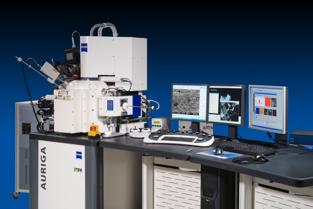
Zeiss Auriga
Field emission scanning electron microscope with ion column, EDX system, EBSD system, and in situ functionality
- SEM point resolution: 1 nm @ 15 kV
- Ion point resolution: 7 nm @ 30 kV
This high resolution SEM is equipped with a multitude of different detectors, suited for most investigations: SE and 4 quadrants BSE detector; inlens SE and inlens ESB detector; SI detector, STEM detector, GIS system for charge compensation; tomographic imaging via slice and stack; sample preparation for TEM;
Tensile and compression testing module for in situ experiments with a 5 kN load cell and heating up to 800 °C

Zeiss EVO 15
Scanning electron microscope (LaB6) with EDS system; imaging with high depth of field; SE detector (Everhart-Thornley) for topography, 5 sectors HDBSD detector for material contrast and crystal orientation; C2DX detector for the extended pressure applications and imaging of samples under fully-hydrated conditions. Sample tilting possible; Point to point resolution 5 nm at 30kV and 10pA;
Extended depth of field and wide field imaging for large samples. Imaging is possible under cryo conditions (down to -170°C) using a cryo holder, under variable pressure (10-4000 Pa depending on mode) and under elevated humidity (up to approx. 100% H2O using a Peltier sample cooling stage) for documentation of hydrated samples.
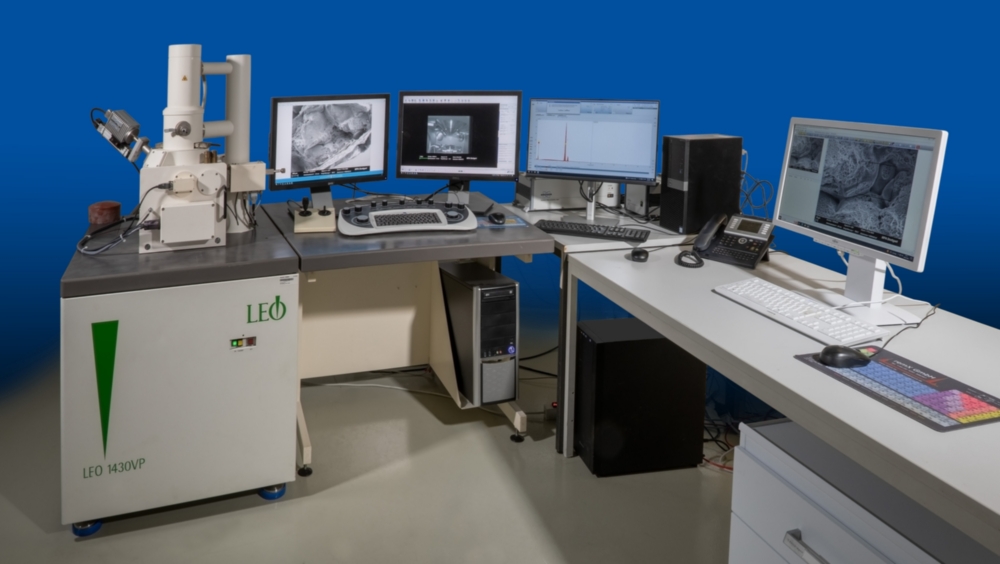
Zeiss Leo 1430 VP
Scanning electron microscope with EDX system; Imaging with high depth of field; high working distance; This system is ideal for specimens with uneven surfaces, like fracture structures
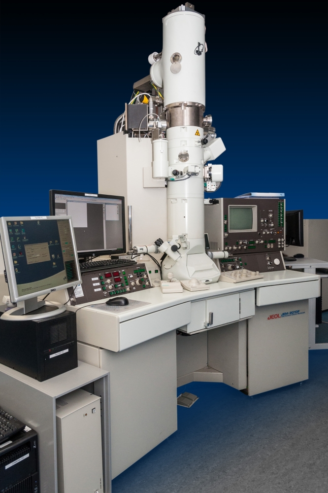
JEOL JSM 2010F
Field emission transmission electron microscope (TEM) with scanning modus and EDX system
- Point resolution 0.2 nm
This TEM is especially suited for the study of precipitates and structures at boundaries as well as dislocation studies. The STEM mode in combination with the EDX system allows for elemental distribution maps with a spatial resolution better than 1 nm. The goniometer can be piezo-controlled for vibration-free specimen positioning.
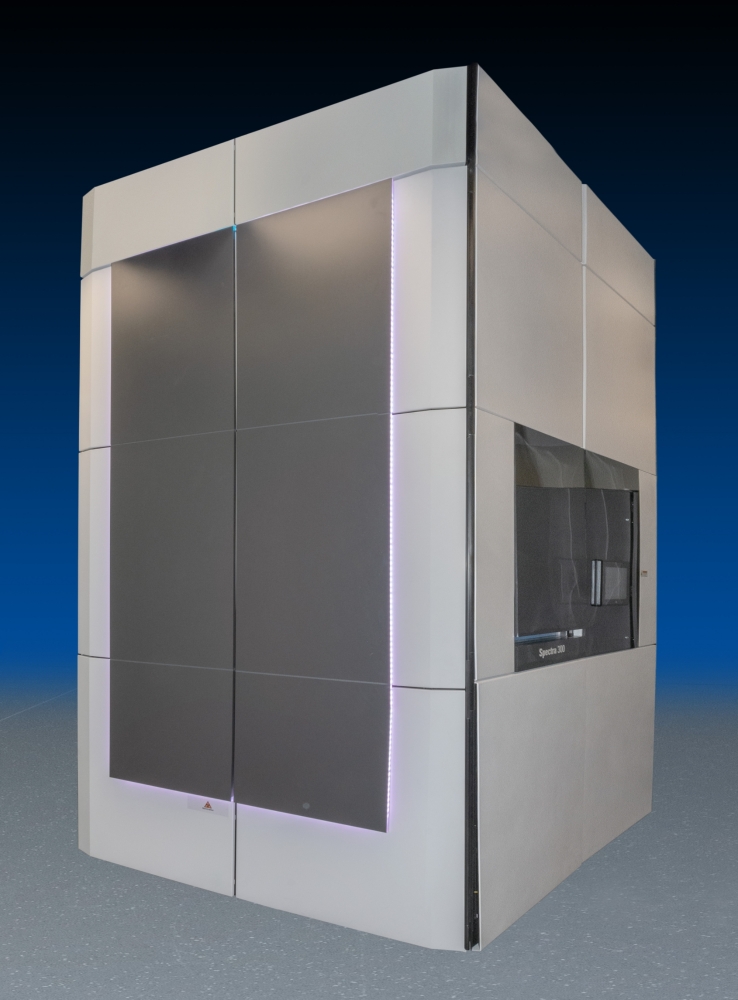
Thermo Scientific Spectra 300
Schottky field emission transmission electron microscope (30-300kV) with Cs S-CORR probe corrector for highest resolution in STEM-mode.
Point resolution: 100 pm in TEM-mode @300 kV or 50 pm in STEM-mode @300 kV or 125 pm in STEM-mode @30 kV). The microscope is equipped with a monochromator (with auto-alignment option) for optimum energy resolution and to control the beam current (e.g. 0.14 nm spot size at 0.3 eV and 0.5 nA @300 kV or 0.14 nm spot size at 0.2 eV and 0.1 nA @ 60 kV).
This high-end TEM is dedicated for STEM and achieves today’s highest specifications. The microscope is equipped with a multitude of detectors and systems that enable quantitative analyses at highest resolution in order to cover a wide range of scientific issues. Beside the BF, ADF, HAADF and iDPC-STEM detectors, there is the 4D-STEM direct electron detector Quantum Merlin which enables ultrafast in situ imaging (up to 2400 Hz) or imaging of very sensitive samples (down to 24 bit/px). For TEM imaging, the microscope uses a fast Ceta 16M camera (4K x 4K at 40 fps and 512x512 at 320 fps). Quantitative analysis is realized by a specimen-tilt independent Super-X EDS-system (<136 eV, 0.7 sr, Fiori-number > 4000) and the EELS Continuum S/1077 spectrometer enabling capture up to 2600 spectra/s in the range of 30-300 kV.
Further equipment is a Gatan Elsa cryo-transfer holder for imaging under cryo conditions + dry pumping station, a tomography holder for tilt series of up to ±70°, an in situ holder (Fusion Select MEMS) for heating and simultaneous electrical measurements, and the NanoMEGAS package enabling precession diffraction and the automatic evaluation of orientation-dependent diffraction patterns
XRCT Nikon X-Tec XTH225
225 kV Microfokus-CT-System
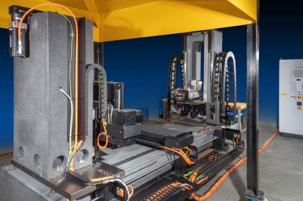
XRCT
180 kV Microfocus CT system; Open, modular, and flexible micro X-Ray Computed Tomography (µXRCT) system with integrated universal testing machine for quasi-static mechanical in situ studies (detail view; front left X-ray source, center testing machine)
Currently in installation: XRCT Procon CT-Alpha Duo
Open computer tomography system with two X-Ray sources:
- 240 kV nano focus tube with minimal focal spot size < 1 µm,
- 320 kV mini focus tube,
- 4K flat panel detector with 100 µm pixelpitch
The CT system will be implemented as open system (without fully radiation safe steel cabinet) in the X-Ray room at the MPA University of Stuttgart. The later implementation of devices for in-situ testing is possible as well as change and addition of X-Ray sources and detector. X-Ray sources and detector are manipulated by motorized axes. Focus-detector-distances between 350 and 1500 mm are possible. Focus-object-distances can be varied between 0.25 and 1400 mm, to reach maximum amplification. The rotary table for CT-scans can handle objects up to 100 kg. 2D radiography is possible even for heavier objects. In addition to X-Ray imaging the CT-system offers the possibility to capture phase contrast and dark field images

Marie-Louise Lemloh
Dr. rer. nat.Manager